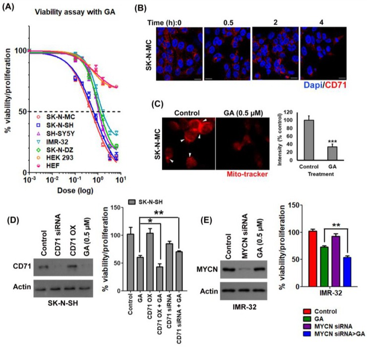
Fig. 2. Gambogic acid preferably targets neuroblastoma cells through CD71. (A) Neuroblastoma cells (IMR-32, SK-N-DZ, SH-SY5Y, SK-N-SH, and SK-N-MC), HEK293 and HEF cells were treated with different (0 to 3.2 µM) doses of GA and the relative percentage of viability with proliferation was calculated by following the formula: % viability, proliferation = (optical density (OD) of the drug-treated sample/OD of the control sample) × 100. The values were plotted with a log dose and the curve-fit was by GraphPad Prism 5. (B) SK-N-MC cells were treated with 0.5 µM of GA, and incubated for different time intervals and fixed. Expression of CD71 was observed using the anti-CD71 antibody. DAPI was used to stain the nuclei. Photographs were obtained by a confocal microscope at 40X magnification. (C) GA-treated SK-N-MC cells were stained with Mito-Tracker Red and imaged under an inverted fluorescence microscope at 40X. (D) GA sensitive SK-N-SH cells were transfected with CD71 overexpression vector or siRNA against CD71 or control vector and with 0.5 µM of GA. Expression of CD71 was confirmed in the transfected cells by Western blot and the relative percentage of viability with proliferation was calculated by MTT assay. The results were expressed as mean ± S.E. (E) MYCN overexpressed IMR-32 cells were transfected with siRNA against MYCN or control siRNA vector with or without GA and Western blot was performed to determine the expression of MYCN; the relative percentage of viability with proliferation was calculated by MTT assay. The results were expressed as mean ± S.E.